Skip to content

MCAT practice test

MCAT - Diagnostic Test - Biology Section

  • 31 questions
  • 42-minute timer
  • Answers and explanations
Embed on your site
  • 31 questions
  • 42 minute timer
  • Every answer explained at the end
  1. Question 1

    The quality of DNA-labeled affinity probes is critical in DNA-assisted protein analyses, such as proximity ligation assays (PLA) and immuno-PCR. Efficient, high-performance methods are therefore required for isolation of pure conjugates from reactions where DNA strands have been coupled to antibodies or recombinant affinity reagents. An important aspect of the synthesis of these molecules is sequentially removing any unconjugated affinity reagents and remaining free oligonucleotides from conjugation reactions, as shown in Figure 1. Figure 1 Illustration of purification of affinity binder-oligonucleotide conjugates First, the conjugation is performed in solution by mixing antibodies with an excess of azide-modified oligonucleotides. The resulting mix containing antibody-DNA conjugates and remaining free antibodies and oligonucleotides is incubated, and the products are immobilized on agarose beads. After washes to remove unconjugated antibodies, the captured antibody-DNA conjugates along with unconjugated oligonucleotides are both released by enzymatic cleavage using the restriction enzyme MlyI that cleaves at the border of the complementary region (Figure 2) between the capture and conjugate oligonucleotides to release single-stranded DNA-coupled conjugates. The cleavage process required both ATP and S-adenosyl-L-met in order to run to completion. The eluate was then captured on protein G beads and washes were conducted to remove any free oligonucleotides with no antibody conjugated. Finally, purified conjugates were eluted from the protein G beads by lowering the pH. These purified high-grade probes extend the capabilities of traditional immunoassays to allow direct detection of proteins and modifications with high specificity and sensitivity. With them, designated protein targets can be readily detected and localized with single molecule resolution and objectively quantified in unmodified cells and tissues. Figure 2 Enzymatic cleavage by MlyI Question 1 In vitro, free oligonucleotides, such as mRNA transcripts, have a short half-life in the cytosol. What mechanism is used to stabilize these molecules?

    Correct answer: A

    The poly-A tail is important for the nuclear export, translation, and stability of mRNA. The tail is shortened over time, and, when it is short enough, the mRNA is enzymatically degraded.
  2. Question 2

    Given the locations of the MlyI cleavage site and recognition sequence, MlyI is NOT a:

    Correct answer: A

    A restriction enzyme is an enzyme that cuts DNA at or near specific recognition nucleotide sequences, known as restriction sites. Type I enzymes cleave at sites remote from the recognition site; they require both ATP and S-adenosyl-L-methionine to function. Since the passage shows a cleavage site very near a recognition site, MlyI must not be a Type I enzyme.
  3. Question 3

    Which nucleophile was likely used to prepare the oligonucleotides used in the purification procedure?

    Correct answer: D

    Paragraph 2 states that the oligonucleotides are azide-modified. Azide (N\(_{3}\)\(^{-}\)) is a powerful nucleophile which can readily add to available electrophiles, such as nucleotide molecules. Content Foundations: Nucleophilicity In organic chemistry, reactions happen when a bond is formed between an electrophile and a nucleophile. Electrophiles are species that ‘want’ more electrons. Often, electrophiles are positively charged or partially positively charged. In contrast, nucleophiles are species that have an excess of electrons, and therefore ‘want’ to use those electrons to form a bond with an electrophile. This is known as nucleophilic attack. Nucleophiles can often be recognized by the presence of at least one free pair of valence electrons, although nucleophilic attack can also take place using electrons from a pi or sigma bond. The factors that make a molecule a strong nucleophile include greater electron density—that is, more lone pairs and, if possible, more charge—and the lack of steric hindrance.
  4. Question 4

    Some proteins and antibodies are susceptible to pH-induced damage. In order to prevent this damage, what would be the most effective additional step to perform at the end of the elution process?

    Correct answer: C

    The passage describes the final step of the process as lowering the pH to elute the proteins off of the beads. To reduce the risk of damage, we need to neutralize any excess acid. 1 M Tris•HCl, pH 8.5 is the buffer that will most effectively dissociate most protein:protein and antibody:antigen binding interactions without permanently affecting protein structure. Eluted protein fractions would best be neutralized immediately by addition of an alkaline buffer. Content Foundations: Acid-base Neutralization In a neutralization reaction, an acid and a base react with each other to produce a salt and water. The actual neutralization that takes place can be thought of as a “canceling out” of each proton from the acid with one hydroxide ion from the base, forming a molecule of H\(_{2}\)O. Despite the name, neutralizations do not always result in a solution of neutral pH (pH = 7 under standard conditions), since the product salt may be acidic or basic. A classic example of an acid-base neutralization reaction is the reaction between HCl (a strong acid) and NaOH (a strong base): HCl(aq) + NaOH(aq) → NaCl(aq) + H\(_{2}\)O(l). The amount of base (or acid) required to fully neutralize an acid (or base) is given by the equation N\(_{acid}\)V\(_{acid}\) = N\(_{base}\)V\(_{base}\), which relates the normalities and volumes of the two solutions. For monoprotic species, where normality and molarity are equal, this simplifies to the fundamental concept of “moles acid = moles base,” while for polyprotic species, it can be described as “moles H\(^{+}\) = moles OH\(^{-}\).” In a titration, complete neutralization occurs at a position termed the equivalence point. The above equation, then, is only valid at the equivalence point, not at any random point over the course of the titration.
  5. Question 5

    Which step of the procedure corresponds to the release of DNA-coupled conjugates?

    Correct answer: C

    We are looking for the step in which the DNA-coupled antibodies are released. Paragraph 2 outlines each step of the procedure shown in Figure 1. Beginning with step A, DNA oligonucleotides are hybridized to the oligonucleotides in the mixture. In step B, agarose beads are used to capture the biotinylated capture oligonucleotides, both in the form of conjugates and free oligonucleotides. In step C, the unconjugated antibodies are removed by washes. In step D, the MlyI enzyme is used to cleave the captured oligonucleotide hybrids, allowing both conjugates and oligonucleotides to be eluted from the solid support. Thus, step D is our answer.
  6. Question 6

    Passage 2 (Questions 6 - 10) The human digestive tract is fundamentally a long tube running through the body, with dedicated sections that are capable of digesting material put in at the proximal end, extracting any useful components from it, then expelling the waste products at the distal end. The small intestine is the site where most chemical digestion is carried out, and where virtually all the absorption of useful materials occurs. The intestines also have a smooth muscle wall with two layers of muscle, which enables the generation of rhythmic contractions that force products of digestion through the intestine. To allow for absorption and secretion, specialized zones known as macula communicans exist between the muscle cells. These directly connect the cytoplasm of two cells, which allows various molecules and ions to pass freely between them. In the large intestine, these connections allow for the movement of ions and fluids. For example, if waste is too solid, it cannot move fluidly and can result in blockage. To prevent this, Cl**\(^{-}\)** ions can be secreted into the lumen. Cations and water will follow the chloride into the lumen by passing through intercellular spaces. This method of transport contrasts typical transcellular transport, where substances travel through the cell membrane. Diseases of the intestines are often due to chronic inflammation, which can arise from imbalances in the regulation of fluids and solute movement. Elevated levels of enzymes like thymidine kinase are often used to predict the risk of colon cancer. In which of the following cell processes does thymidine kinase most likely play a role?

    Correct answer: B

    The passage states that thymidine kinase is used to predict cancer activity. Cancer is a disease categorized by rapid, unregulated cell growth and replication. The name "thymidine" (a nucleotide base) also indicates that the enzyme has a key role in DNA synthesis and therefore in cell division. We’re also expected to know that the "kinase" part of the name indicates that the enzyme catalyzes the addition of a phosphate group to its substrate. While knowledge about specific diseases is generally not required for MCAT success, there are notable exceptions to this rule. One such exception is cancer, which can be summarized as abnormal gene expression. To make sense of this statement, let’s review the steps involved in cancer development, or oncogenesis. The term “tumor” describes any abnormal proliferation of cells. Benign tumors remain localized, whereas malignant tumors (which are what the term “cancer” properly refers to) can invade other organs and tissues in the body in a process called metastasis. The first step in oncogenesis, tumor initiation, involves changes that allow a single cell to proliferate abnormally. This means that the cell must develop the ability to bypass regulatory steps of the cell cycle that normally help to limit mitotic proliferation. Tumor progression occurs as a cell develops the ability to proliferate even more aggressively, such that its descendants are selected for and come to predominate the growing tumor. In addition, malignant cells often undergo mutations that promote their own growth and the development of blood vessels to feed them (angiogenesis). Oncogenesis is most often associated with mutations that occur by random chance (and elude the normal DNA repair machinery in the cell) or as a result of mutagenic compounds known as mutagens or carcinogens. (Examples of mutagens include ultraviolet light and certain chemicals, such as reactive oxygen species.) These mutations alter the functionality of crucial genes in the cell. However, oncogenesis is also associated with dysregulation of gene expression, as the abnormally elevated expression of genes involved in growth and proliferation can help contribute to the development of a tumor. The genes involved in oncogenesis can be divided into two groups: oncogenes and tumor suppressor genes. The basic difference between them is that oncogenes function to promote abnormal growth and proliferation, leading to cancer, while tumor suppressor genes function to prevent tumorigenic properties. Oncogenes can arise from the mutation of other genes, termed proto-oncogenes. If not mutated, proto-oncogenes do not promote cancer, but certain mutations or inappropriately elevated gene expression can effectively turn them into oncogenes.
  7. Question 7

    Chitin’s flexibility and strength make it favorable as a biodegradable surgical thread. All of the following describe the portion of a chitin molecule shown below EXCEPT:

    Correct answer: B

    Chitin (C\(_{8}\)H\(_{13}\)O\(_{5}\)N)n is a long-chain polymer of a N-acetylglucosamine, a derivative of glucose. The molecule shown comprises two of these rings attached via a β-1,4 linkage. As such, it can be classified as a carbohydrate, a polymer, and a disaccharide. It does not contain amino acid monomers joined via amide bonds, so it cannot be classified as a polypeptide. Content Foundations: Biomolecules There are four main classes of biomolecules: amino acids/proteins, lipids, carbohydrates, and nucleic acids. All of these classes except for lipids can occur as polymers, as proteins can be seen as polymers of amino acids, DNA and RNA are polymers of nucleic acids, and compounds such as starch, cellulose, and glycogen are polymers of carbohydrates. Amino acids contain a central carbon to which –NH\(_{2}\), –COOH, –H, and –R groups are added, and they combine by peptide bonds to form dipeptides, tripeptides, oligopeptides, and proteins. The key functionality of an amino acid is determined by its side chain (–R). Proteins are the building blocks of the body, and are involved in structural and signaling roles. Lipids are nonpolar (or amphipathic, with both polar and nonpolar areas) molecules that play roles in signaling, structural functions, and energy storage. The main categories of lipids that you must know for the MCAT are fatty acids and the derivatives thereof (triacylglycerols, phospholipids, and sphingolipids), cholesterol and its derivatives (steroid hormones and vitamin D), prostaglandins, and terpenes and terpenoids. Carbohydrates are important for energy storage and are used in metabolism. They are made up of carbon, hydrogen, and oxygen, often in the ratio C\(_{x}\)(H\(_{2}\)O)\(_{y}\). Important monosaccharides include glucose, fructose, and galactose. Key disaccharides are sucrose (glucose + fructose), lactose (glucose + galactose), and maltose (glucose + glucose). Polysaccharides (starch and glycogen) are polymers of glucose that are used for energy storage in plants and animals, respectively. Nucleic acids are involved in the storage and transmission of biological information. They are made up of nucleotides, which have three components: a nitrogenous base, a five-carbon sugar (ribose in RNA and deoxyribose in DNA), and at least one phosphate group. RNA contains uracil (U) instead of thymine (T) and is generally single-stranded, whereas DNA is generally double-stranded. DNA is used for the long-term storage of genetic information, while RNA is used for gene expression.
  8. Question 8

    Patients with excess fat are more likely to require larger therapeutic doses of which vitamin?

    Correct answer: C

    The lipid-soluble vitamins are A, D, E, and K. The water-soluble vitamins are B and C. Greater amounts of subcutaneous fat sequester more of the lipid-soluble vitamins and lower their release into the circulation. Thus, excess fat increases the initial dose of vitamin required to achieve a particular effect. Content Foundations: Vitamins, Minerals, Cofactors, and Coenzymes An enzyme (a biological catalyst) may require another chemical compound to be present in order for it to carry out its biological functionality. In general, such “helper” molecules are known as cofactors. Cofactors can be either inorganic (with some common examples including metal ions such as Mg\(^{2+}\), Zn\(^{2+}\), and Cu\(^{+}\)) or organic, and organic cofactors are sometimes known as coenzymes. Many coenzymes are vitamins or derivatives of vitamins, and they often contribute to the function of enzymes by carrying certain functional groups from one place to another in a reaction. Perhaps the most well-known example of this is coenzyme A, which transfers acyl groups from one place to another. Coenzymes that are tightly (or even covalently) bonded to their enzyme are known as prosthetic groups. A famous example of an organometallic prosthetic group is heme, which contains an iron ion in the center of a porphyrin ring, and is attached to oxygen-transport proteins such as hemoglobin and myoglobin. Vitamins are non-macronutrient compounds that are vital for healthy functioning and cannot be synthesized in adequate quantities by the body, meaning that they must be obtained from external sources. The B vitamins and vitamin C are coenzymes used for various important reactions, and are water-soluble. Vitamins A, D, E, and K are lipid-soluble. Vitamin A plays a key role in vision, vitamin D in calcium and phosphate absorption from the gastrointestinal tract, vitamin E is an antioxidant, and vitamin K promotes coagulation. Like vitamins, minerals contribute to essential roles in the body and must be obtained from the diet, but unlike vitamins, minerals are inorganic. The most important minerals in the body are calcium, phosphorus, magnesium, sodium, and potassium.
  9. Question 9

    Which of the following will cause a blood pH of 8.2?

    Correct answer: D

    Content Foundations: Bicarbonate Buffering Acid-base buffers confer resistance to a change in the pH of a solution when hydrogen ions or hydroxide ions are added or removed to solution. An acid-base buffer typically consists of a weak acid and its conjugate base. The most important buffer to know for the MCAT is the bicarbonate buffer system, which is shown below. H\(_{2}\)O(aq) + CO\(_{2}\)(g) ↔ H\(_{2}\)CO\(_{3}\)(aq) ↔ H\(^{+}\)(aq) + HCO\(_{3}\)**\(^{-}\)*(*aq) Carbonic acid (H\(_{2}\)CO\(_{3}\)) has the conjugate base of HCO\(_{3}\)**\(^{-}\)**. Buffers work because the concentrations of the weak acid and its salt are large compared to the number of protons or hydroxide ions added or removed. When protons are added to the solution from an external source, some of the bicarbonate in the buffer is converted to carbonic acid, using up the protons added; when hydroxide ions are added to the solution, protons are dissociated from some of the carbonic acid in the buffer, converting it to bicarbonate and replacing the protons lost. Buffers resist pH changes best when the pH values are at or near the pK\(_{a}\) value for the acid/base used, because that is when the conjugate acid and base have equal concentrations. Optimal buffering occurs when the pH is within approximately 1 pH unit from the pK\(_{a}\) value of the system. The pH of a buffer solution can be calculated using the Henderson-Hasselbalch equation: pH = pK\(_{a}\) + log [conjugate base] / [acid]. On Test Day, you may see the bicarbonate buffer concept tested from a chemical, biochemical, or biological perspective. For example, hyperventilation (rapid shallow breathing) results in excess CO\(_{2}\) being expelled from the blood, causing the pH to rise. In response, the buffer needs to release more H\(^{+}\) to lower the pH back to physiological norms. An additional fact to be aware of is that other mechanisms in the body are also used to regulate pH, since carbonic acid works best at a pH below physiological conditions, because its pK\(_{a1}\) (pK\(_{a1}\) = 6.3, pK\(_{a2}\) = 10.3) is much lower than the normal pH of blood (7.4).
  10. Question 10

    The data in the passage suggest that the level of CWP begins to drop most quickly during which time period of encystation?

    Correct answer: B

    Figure 1 shows the up- or down-regulation of cwp genes over two-hour intervals. The 4-6 hpie time period shows the largest down-regulation, 15%, as evidenced by the gray bar on the lower half of the plot.
  11. Question 11

    With which of the following do the arginine side chains found on histone proteins most likely have the strongest interactions?

    Correct answer: C

    Arginine is a basic amino acid, meaning that it is positively charged at physiological pH. Positive species interact best with negative species, and of the answers listed, only phosphate groups are negatively-charged. Content Foundations: Biomolecules There are four main classes of biomolecules: amino acids/proteins, lipids, carbohydrates, and nucleic acids. All of these classes except for lipids can occur as polymers, as proteins can be seen as polymers of amino acids, DNA and RNA are polymers of nucleic acids, and compounds such as starch, cellulose, and glycogen are polymers of carbohydrates. Amino acids contain a central carbon to which –NH\(_{2}\), –COOH, –H, and –R groups are added, and they combine by peptide bonds to form dipeptides, tripeptides, oligopeptides, and proteins. The key functionality of an amino acid is determined by its side chain (–R). Proteins are the building blocks of the body, and are involved in structural and signaling roles. Lipids are nonpolar (or amphipathic, with both polar and nonpolar areas) molecules that play roles in signaling, structural functions, and energy storage. The main categories of lipids that you must know for the MCAT are fatty acids and the derivatives thereof (triacylglycerols, phospholipids, and sphingolipids), cholesterol and its derivatives (steroid hormones and vitamin D), prostaglandins, and terpenes and terpenoids. Carbohydrates are important for energy storage and are used in metabolism. They are made up of carbon, hydrogen, and oxygen, often in the ratio C\(_{x}\)(H\(_{2}\)O)\(_{y}\). Important monosaccharides include glucose, fructose, and galactose. Key disaccharides are sucrose (glucose + fructose), lactose (glucose + galactose), and maltose (glucose + glucose). Polysaccharides (starch and glycogen) are polymers of glucose that are used for energy storage in plants and animals, respectively. Nucleic acids are involved in the storage and transmission of biological information. They are made up of nucleotides, which have three components: a nitrogenous base, a five-carbon sugar (ribose in RNA and deoxyribose in DNA), and at least one phosphate group. RNA contains uracil (U) instead of thymine (T) and is generally single-stranded, whereas DNA is generally double-stranded. DNA is used for the long-term storage of genetic information, while RNA is used for gene expression.
  12. Question 12

    Given passage information, Giardia most likely transports which of the following proteins to the extracellular surface?

    Correct answer: A

    According to paragraph 2, the CWM is "composed of at least 3 cyst wall proteins (cwp1-3)," one of which is cwp3. The CWM is eventually attached to the intestinal cell membrane that the parasite inhabits. This suggests that there must be a mechanism for transporting this protein from the interior of the parasitic cell to the host cell membrane. Content Foundations: Histones and DNA Packaging The human genome contains approximately 3 billion base pairs. To fit into human nuclei, which are generally about 6 μm (6 × 10\(^{-6}\) m) in size, DNA must be compressed. Subdividing the genome into linear chromosomes accomplishes some of this task, but the rest of the job is done by histones and chromatin. Histones are proteins that act as spools for DNA to wind around. They are composed of various subunits known as H1, H2A, H2B, H3, and H4. The core of a histone contains two dimers of H2A and H2B and a tetramer of H3 and H4, while H1 serves as a linking unit. Approximately 200 base pairs of DNA can be wound around a histone, and the complex formed by DNA and a histone is known as a nucleosome. The phrase “beads on a string” is often associated with the appearance of nucleosomes under electron microscopy, and chromatin refers to the structure formed by many nucleosomes. Two distinct forms of chromatin exist: euchromatin and heterochromatin. Euchromatin is a loose configuration that is difficult to see under light microscopy and allows DNA to be readily transcribed. Throughout interphase (i.e., most of the cell cycle), DNA generally exists as euchromatin. Heterochromatin is the tightly coiled, dense form of chromatin that is visible during cell division and is present to a lesser extent even during interphase. On a biochemical level, charge plays a major role in the interactions between histones and DNA. Histones are highly alkaline and are positively charged at physiological pH, which facilitates their interaction with the highly negatively charged phosphate groups on the backbone of DNA. Modifications like acetylation of histones reduce that positive charge, making histones interact with DNA less closely, which in turn facilitates transcriptional activity.
  13. Question 13

    The functions of the ESVs of Giardia most closely resemble what cell organelle?

    Correct answer: B

    Paragraph 2 tells us that encystation specific vesicles (ESVs) give the organism the ability to modify or partition cyst wall cargo during secretory transport. This is most similar to the functions of the Golgi apparatus, which packages and modifies cellular material for transport. Eukaryotic cells contain several membrane-bound organelles. The three most commonly-tested organelles on the MCAT are the nucleus, mitochondria, and ribosomes. The nucleus contains DNA and a sub-organelle known as the nucleolus, which is responsible for ribosome assembly. Mitochondria are sometimes known as the powerhouse of the cell; they are self-replicating organelles with their own DNA that are the location of the citric acid cycle and oxidative phosphorylation in eukaryotes. Ribosomes themselves are not membrane-bound organelles, and they are not unique to eukaryotes (although prokaryotic ribosomes have a different structure). They play the crucial role of protein translation. Once proteins are synthesized, the Golgi apparatus modifies them and packages them into membrane-bound vesicles that are then sent to the ultimate destination of the proteins. In addition, the endoplasmic reticulum is a net-like organelle that extends out from the nuclear membrane. It is divided into the rough ER and the smooth ER. The rough ER is rough because it is covered with ribosomes, which are the site of protein synthesis. In contrast, the smooth ER does not have ribosomes, and is involved in lipid metabolism (both synthesis and breakdown), the production of steroid hormones, and detoxification. Lysosomes are the garbage disposal system of the cell; material from outside the cell enters the lysosomes through endocytosis, while material from inside the cells enters through autophagy. They contain hydrolases that operate best at acidic pH levels, and the lysosomes are therefore kept at a pH of 4.5-5.0. Finally, peroxisomes play a major role in the metabolism of very-long-chain lipids by breaking them down to medium-chain lipids that are transported to the mitochondria for further processing and play a role in detoxification of substances such as ethanol.
  14. Question 14

    Passage 5 (Questions 24 - 28) Giardiasis, caused by the intestinal flagellate protozoa Giardia lamblia, is a common intestinal disease. Giardia are unique in their possession of two nuclei that are similar in appearance, DNA content, and transcription. Usually G. lamblia are transmitted by water contaminated with cysts. G. lamblia have seven complex genotypes, but only two genotypes cause human infections. The illness is transmitted by differentiation into infective cysts. Three known cyst wall proteins (CWPs) are synthesized in a concerted manner during differentiation into cysts. Encystation specific vesicles (ESVs) give the organism the ability to modify or partition cyst wall cargo during secretory transport. These organelles aid the accumulation and maturation of cyst wall material (CWM) composed of at least 3 cyst wall proteins (cwp1-3) complexed with β-(1-3)-N-acetyl-D-galactosamine polymer. CWPs are eventually deposited in the plasma membrane of the host’s intestinal cells. Trafficking of mature CWM from ESVs to the cell surface, where it forms the cyst wall, is rapid. During cyst formation, a round of DNA replication occurs. Then, nuclear replication takes place, with the DNA divided equally among the resulting 4 nuclei, followed by a second round of DNA replication. Figure 1 Regulation of cwp abundance within 2-hour transitions during the 14-hour encystation time-course (hpie = hours post initiation of encystations. N = total protein in sample) At any given time throughout the parasite's lifespan, only one cwp gene is expressed. During an infection, however, the particular cwp gene that is transcribed regularly alternates in a process termed antigenic variation. Both expressed and silent cwp genes are located in the nuclear periphery, but in different regions. In expressed cwp genes, promoter histone H4 is acetylated on arginine 6 and trimethylated on arginine 2; in contrast, silent cwp genes include a trimethylated histone H4 at arginine 6. During the homologous recombination in cyst nuclei, type II topoisomerases assist in chromosome replication and tissue development for Giardia differentiation into dormant cysts. In addition, the levels of cyst formation and the cwp1-3 gene expression are increased by Topo II overexpression.

    Correct answer: C

    he protozoa will typically be diploid, and as stated in the second paragraph, cyst formation involves a round of DNA replication, followed by nuclear replication, followed by a second round of DNA replication. This process is illustrated below, and the presence of four 4n nuclei yields a final total ploidy of 16n. Content Foundations: Ploidy, Aneuploidy, and Nondisjunction Ploidy refers to how many copies of each chromosome a cell has. In humans, the vast majority of cells are diploid (2n), meaning that they contain two copies of each chromosome (except for the sex chromosomes; females have two X chromosomes and males have an X and a Y chromosome). Such cells are known as somatic cells—that is, the cells of the body. In contrast, germ cells (i.e., ova and spermatozoa) are haploid (n), meaning that the only have a single copy of each chromosome. Aneuploidy results from having too many or too few copies of a given chromosome. This results from nondisjunction in anaphase during cell division. Having only one copy of a chromosome is known as monosomy, and having three copies is known as trisomy. Aneuploidy is commonly discussed as occurring in meiosis, and indeed, this is the only way for aneuploidy to be inheritable. For this reason, nondisjunction during meiosis is the cause of aneuploidies such as Down syndrome (trisomy 21) or Turner syndrome (monosomy X). However, nondisjunction during mitosis can also occur, and this is extremely common in cancer cells.
  15. Question 15

    Given the findings in the passage, which additional result would MOST strongly indicate that TGFBR1 and TGFBR2 regulate TGFβ-dependent LEC proliferation?

    Correct answer: B

    Experiment 1 of the passage shows that treatment with TGFBR1 and TGFBR2 antagonists led to lower levels of epithelial cell production. In order to strengthen this conclusion, we want to show a positive correlation between TGFBR activity and epithelial proliferation.
  16. Question 16

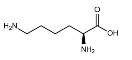
    The structure of lysine is shown below. This molecule is best described as:

    Correct answer: B

    The structure of lysine is dominated by a carboxylic acid group (R(CO)OH) and two basic amine (NH\(_{2}\)) groups. Therefore, it is best described as polar and basic.
  17. Question 17

    After injury-induced cataract formation has begun, which of the following are LEAST likely to be found in nearby monocytes?

    Correct answer: C

    Tight junctions are areas where the membranes of two closely-joined cells adhere tightly and form a near-impenetrable barrier. This is a hallmark of epithelial barriers and is not likely to be a property of monocytes, which are immune cells. Content Foundations: Cell Junctions Cell junctions describe ways in which neighboring cells interact with each other to form structures. There are three main types of cell junctions that you should be aware of: anchoring junctions, gap junctions, and tight junctions. Anchoring junctions include adherens junctions, which are associated with cadherins. The essential idea behind anchoring junctions is that they connect cytoskeletal components of the cell with other cells and/or the extracellular matrix, thereby contributing to the overall structural stability of tissues. Adherens junctions involve cadherin-mediated connections between actin filaments and other cells and the extracellular matrix. Desmosomes also involve cadherin, but in this case cadherin connects intermediate filaments to other cells. Hemidesmosomes are junctions in which integrins connect the intermediate filaments of cells to the extracellular matrix. Gap junctions are formed by connexin proteins, which connect cells in a way such that diffusion can take place between them, enabling communication, without involving direct contact between the cytoplasmic fluids of each cell. Gap junctions are relatively less common, but they play certain crucial roles within the body. The most important example for the MCAT is in cardiac muscle, where gap junctions allow cells to contract at the same time. Finally, tight junctions are found in epithelial cells. As the name suggests, the cells in tight junctions are linked very closely to each other, preventing solutes from being able to move freely from one tissue into another. A classic example is the blood-brain barrier, where the epithelial cells in blood vessels in the brain form very tight junctions that allow the close regulation of which substances from the bloodstream can enter the central nervous system. Some types of epithelial tissue have relatively few tight junctions; these are known as leaky epithelia. Examples include some parts of the kidney.
  18. Question 18

    Monocytes, in conjunction with epithelium-derived factors, can act to facilitate which biological process?

    Correct answer: D

    Monocytes are the largest type of white blood cells. Factors secreted by the epithelium help form a cascade that leads to accumulation of immune cells at the site of an injury.
  19. Question 19

    Passage 4 (Questions 19 - 23) Cataract generation triggered by lens injury is considered to be driven by the action of the proliferation and epithelial-mesenchymal transition (EMT) of lens epithelial cells (LECs) as a complication of injury and inflammation. The epithelium quickly expands, creating a hundredfold increase in the number of cells. In response to trauma, irritation, or surgery, cytokines and growth factors increase in the aqueous humor and stimulate LECs to proliferate and undergo EMT. This response is significantly diminished in organisms where EMT is inhibited by the inactivation of transforming growth factor β (TGFβ), especially TGFβ2, the major isoform in the aqueous humor. During the process of EMT, LECs undergo cytoskeletal rearrangement, with the addition of a large amount of extracellular matrix proteins, such as collagen and fibronectin. In anterior cataracts, the proliferation and EMT of LECs in situ leads to the formation of opaque plaques just beneath the lens anterior capsule. The production of TGFβ is regulated by histone deacetylase-1 (HDAC1). HDAC1 removes acetyl groups from N-acetyl lysine groups on histone. Once synthesized, TGFβ is secreted by activated monocytes in the aqueous humor. Since monocytes mediate almost 80 percent of local TGFβ concentrations, researchers tested the theory that TGFβ is responsible for monocyte impact on LEC production. The number of LECs expressing MIB1, a nuclear protein, was utilized as a measure of LEC proliferation and expected EMT. Experiment 1 After simulated injury, monocyte/LEC cultures were treated with an antibody that lyses all but a negligible fraction of available monocytes; a division of these cultures was also treated with a TGFβ agonist. A second set of cell cultures was treated with antagonists of epithelial cell TGFβ receptors TGFBR1 and TGFBR2 (Table 1). Table 1 Results of the study on monocyte lysis, TGF agonist, and TGF receptor antagonists Experiment 2 In an effort to determine whether MIB1 is associated with LEC regulation, LEC density was assessed during the course of epithelial growth in wild-type and MIB1-null cell cultures (Figure 1).Figure 1 Growth of LECs after an injury in both wild-type and MIB1-null strains HDAC1 MOST likely plays a direct role in:

    Correct answer: A

    Paragraph 3 states that HDAC1 regulates the production of TGFβ and acts to remove acetyl groups from lysine residues of histones. This is important to gene regulation because DNA is wrapped around histone proteins, and DNA expression is regulated by the addition and removal of acetyl groups. Deacetylation attracts DNA to histones more tightly, inhibiting transcription. In summary, histone acetylation generally serves to increase gene expression, while deacetylation, along with DNA methylation, acts to silence gene expression. Content Foundations: Gene Expression The central dogma of molecular biology states that information is passed from DNA to RNA to protein. This means that when a cell needs more of a certain protein, it can increase the degree to which the gene corresponding to that protein is transcribed. Transcribing more or less of a gene in response to the cell’s needs is known as gene expression. It plays a major role in the differentiation of organs in multicellular organisms, and can also vary on shorter time scales in response to changing environmental conditions. The details of how gene expression is regulated in eukaryotes are quite intricate, but you should be aware of some key concepts for the MCAT. Promoters are regions of DNA that lie upstream to a given gene and initiate transcription by binding specific transcription factors that contribute to the binding of RNA polymerase. Additionally, expression is upregulated by enhancers, which are DNA sequences that can be located further from the gene of interest, and work by binding transcription factors that twist DNA into a hairpin loop, bringing distant regions into close proximity for transcription to begin. Silencers are the opposite of enhancers in eukaryotic cells; they are regions of DNA to which transcription factors known as repressors bind. Additionally, the methylation of C and A residues can reduce transcription. Methylation is associated with epigenetics, which refers to inheritable phenotypic changes involving mechanisms other than the alteration of the genome itself. Gene expression can also be regulated on the level of nucleosomes (i.e. chromatin and histones). Acetylation promotes transcription by attaching acetyl groups to lysine residues on histones, making them less positively-charged and causing a looser wrapping pattern that allows transcription factors to access the genome more easily. Finally, non-coding RNA plays a role in gene expression. MicroRNA (miRNA) strands are single-nucleotide strands incorporated into an RNA structure with a characteristic hairpin loop, while small interfering RNA (siRNA) molecules are short and double-stranded. Both tend to be approximately 22 nucleotides in length, and silence genes by interrupting expression between transcription and translation.
  20. Question 20

    A molecule enters a cell and creates pores in the inner mitochondrial membrane. How will ATP generation as a result of oxidative phosphorylation be affected?

    Correct answer: D

    The energy released by oxidative phosphorylation and used to make ATP is generated from potential energy in the form of an H\(^{+}\) gradient, which creates an electrochemical gradient across the mitochondrial membrane. If this membrane is disrupted, this gradient cannot be maintained. Without this gradient, oxidative phosphorylation cannot continue to produce ATP. Content Foundations: Oxidative Phosphorylation The final major process of aerobic respiration is oxidative phosphorylation, through which the majority of aerobically-derived ATP is synthesized. This process begins by passing electrons through a series of chemical reactions, known as the electron transport chain (ETC), to a final electron acceptor, oxygen. This is the only time in eukaryotic aerobic respiration where oxygen is directly required. These reactions take place in specialized proteins where the energy from NADH and FADH\(_{2}\) is used up, molecular oxygen is reduced into water, and approximately 30-36 ATP are created from ADP and inorganic phosphate. The electron transport chain (ETC) uses free oxygen as the final electron acceptor of the electrons removed from NADH and FADH\(_{2}\) formed in glycolysis and the Krebs cycle (also known as the citric acid cycle). The ETC is composed of four large protein complexes (Complexes I-IV) embedded in the inner mitochondrial membrane and two small electron carriers shuttling electrons between them. Complex I is known as NADH dehydrogenase, II is known as succinate dehydrogenase, III is known as cytochrome bc or c, and IV is known as cytochrome c oxidase. Electrons are released from NADH and FADH\(_{2}\) through a series of reactions. In the ETC, the energy released from the series of electron transfers is used to pump H\(^{+}\)across the membrane. The unequal concentrations of H\(^{+}\) ions across the membrane establishes an electrochemical gradient, leading to chemiosmosis, or the passive diffusion of the protons down their concentration gradient, which is coupled to ATP synthase. This proton movement generates 90% of the ATP synthesized during oxidative phosphorylation. The electrons passing through the electron transport chain gradually lose energy until eventually they are donated to O\(_{2}\), which accepts two H\(^{+}\)ions and is transformed into water. If the proton gradient is disrupted or destroyed, chemiosmosis can become uncoupled from the ETC, resulting in little to no ATP generation despite the transfer of electrons carrying on. Many poisons and toxins act by uncoupling the proton gradient from ATP synthase.
  21. Question 21

    Which of the following is an element of humoral immunity?

    Correct answer: B

    Humoral immunity is part of the body’s adaptive immune response. Humoral immunity is provided by B cell activity, which promotes antibody (immunoglobulin) responses. Antibodies can recognize polysaccharide, phospholipid, and nucleic acid antigens to help the body fight extracellular bacteria, viruses, and toxins. Content Foundations: Immune System The immune system refers to the complex set of mechanisms that the body uses to protect itself against foreign invaders and malfunctioning cells originating from the body itself. The highest-level distinction in the immune system is between the innate (or non-specific) immune system, which responds generally to threats but does not learn to recognize specific foreign bodies/molecules, and the adaptive immune system, which does. The non-cellular component of the innate immune system includes anatomical barriers and signaling molecules such as cytokines and complement proteins, while the cellular component includes a range of white blood cell types (leukocytes) that play various roles in responding to threats. White blood cells include neutrophils, lymphocytes, monocytes (which differentiate into macrophages or dendritic cells), eosinophils, basophils, and mast cells. The various components of the innate immune system can act independently or be coordinated in the process of inflammation. The adaptive immune system includes B cells and T cells, both of which are lymphocytes that are produced in the bone marrow and mature in the lymphatic system. B cells recognize antigens and secrete large amounts of antibodies in response. The human body utilizes five classes of antibodies: immunoglobulin (Ig) A, IgD, IgE, IgG, and IgM, which differ in the details of their heavy chains. This response is known as humoral immunity. In contrast, T cells correspond to the cell-mediated branch of the adaptive immune system. T cells recognize cells that were originally self, but have been damaged by viral infections or have malfunctioned in ways likely to turn them into cancer cells. Then, various subgroups of T cells either directly attack compromised/foreign cells or mobilize responses to them based on antigen fragments that are presented by major histocompatibility complex (MHC) class I and II.
  22. Question 22

    Questions 16-18 do not refer to a passage and are independent of each other. Question 16 Which of the following is MOST likely to use a protein channel to cross the eukaryotic cell membrane?

    Correct answer: B

    The eukaryotic cell membrane is composed of a phospholipid bilayer, cholesterol, and various transmembrane protein pores, channels, and gates. The largely hydrophobic nature of the lipid bilayer prevents ions and polar solutes from diffusing across the membrane, while allowing for the free movement of hydrophobic molecules. Calcium ions are charged, and so would be unable to pass through the membrane in significant amounts. Thus, calcium ions require an ion channel. Content Foundations: Membrane Transport The plasma membrane of eukaryotic cells is primarily composed of a lipid bilayer of amphipathic phospholipids with hydrophilic heads and hydrophobic tails. It also contains cholesterol and membrane proteins. Transmembrane proteins are membrane-spanning proteins with hydrophilic cytosolic and extracellular domains and a hydrophobic membrane-spanning domain. Additionally, peripheral proteins are only transiently attached to integral proteins or peripheral regions of the lipid bilayer, and lipid-anchored proteins are covalently bound to membrane lipids without actually contacting the membrane directly. Membrane transport is accomplished through several mechanisms. Some molecules, like small gases, can directly diffuse through the membrane. This is known as simple diffusion, and is an example of passive transport because no energy is necessary. Osmosis is a type of simple diffusion in which water moves in or out of the cell to attempt to equalize concentrations of solute. Facilitated diffusion is another form of passive transport where no energy is necessary because molecules diffuse down their concentration gradient, but a transmembrane channel is necessary because the molecule may be too large or polar for simple diffusion. Ions are often transported through facilitated diffusion, and aquaporins are facilitated diffusion channels for water that augment osmosis. In primary active transport, energy is used directly to move a solute against its gradient through a transmembrane channel. Secondary active transport is a more complicated system in which the energy stored in an electrochemical gradient established via primary active transport is used to facilitate the movement of a solute. An example is the sodium-calcium exchanger, which allows three Na\(^{+}\) ions to flow down their concentration gradient, which was previously established by a primary active transport mechanism, into the cell, while transporting one Ca\(^{2+}\) ion out. Endocytosis is used to ingest larger materials. It is divided into pinocytosis and phagocytosis. In pinocytosis, cells engulf liquid substances, while in phagocytosis, they engulf solid particles. The basic pathway of endocytosis involves recognition of a target molecule at the plasma membrane, followed by invagination and the formation of a vesicle on the inside of the cell. Exocytosis can be thought of as endocytosis in reverse. It is used to release hormones, neurotransmitters, membrane proteins and lipids, and other materials.
  23. Question 23

    The symptoms of botulism took 12-18 hours to be observed in the patient after exposure was confirmed. How is this delay best explained?

    Correct answer: A

    If growing bacteria produce the toxin, then it will take time for the bacteria to generate sufficient quantities of the toxin to cause illness.
  24. Question 24

    Which of the following is NOT a function of the sympathetic nervous system?

    Correct answer: B

    Content Foundations: Sympathetic and Parasympathetic Nervous Systems The autonomic nervous system, which controls involuntary responses in the body, including things like sweating, blushing, and pupil dilation, is divided into the sympathetic and parasympathetic systems. The sympathetic nervous system stimulates the body in the classic “fight or flight” response, mediated by hormones such as epinephrine and norepinephrine. If the body needs to get ready for action, it will dilate the pupils, raise the heart rate, and increase blood flow to skeletal muscles to prepare for sudden action. By contrast, the parasympathetic nervous system is the “rest and digest” system that increases blood flow to the digestive system, slows the heart rate, constricts the pupils, and generally exerts actions opposite to those of the sympathetic nervous system. In turn, the autonomic nervous system is part of the peripheral nervous system, which describes the nervous system throughout the body and is distinguished from the central nervous system, which corresponds to the brain and spinal cord. The peripheral nervous system is also divided into the visceral nervous system, which modulates the digestive system, and the somatic nervous system, which connects to skeletal muscle to allow for voluntary movement.
  25. Question 25

    Which molecule could be used to specifically detect toxin D in the cultures described in the passage?

    Correct answer: A

    To recognize the antigens on the botulinum protein, an antibody could be used. Antibodies are made to recognize and bind specific epitopes of biomolecules like proteins.
  26. Question 26

    The easiest method to separate the two subunits of the botulinum protein for subsequent analytical purposes would be:

    Correct answer: D

    The second paragraph states that the two subunits are different in size (100 kDa vs. 50 kDa). Size-exclusion chromatography could therefore be used to separate the two proteins by size. Content Foundations: Separations Separation techniques are widely used in organic chemistry to prepare purified substances for analysis or reaction. The best technique for a given scenario often depends on the phases of the substances being separated. If all are liquids, one may be able to utilize distillation, which aims to separate liquids by utilizing the difference between their boiling points. The liquids are initially held in the same round-bottom flask, termed the distilling flask. This flask is positioned above a heat source, typically a Bunsen burner. The top of the flask is connected to a column, which leads to a downward-sloping glass condenser over a receiving flask. The condenser is held within a glass casing through which cold water is pumped. As the round-bottom flask is heated, the liquid with the lower boiling point will begin to vaporize, and its vapor will travel up the column and re-condense to fall into the receiving flask. The eventual result is a receiving flask that is rich in the lower-BP liquid, while the distilling flask will still contain the liquid(s) with the higher BP. If boiling points are very high, a vacuum may also be used to lower atmospheric pressure, which lowers the boiling points of all substances involved. Recrystallization is used to purify a solid product that contains impurities. This process involves the dissolution of the solid in a solvent and subsequent heating. The solid then dissolves and is cooled, causing it to solidify (crystallize) again. As the lattice structures of solids tend to exclude impurities, each subsequent recrystallization results in a progressively purer compound. Chromatography is a broad set of separatory techniques based on relative affinity, or tendency for a compound to attract to a certain solvent or structure. Specifically, sample molecules vary in their affinities for a mobile (moving, typically solvent-based) phase versus a stationary (static) phase. In column chromatography, the stationary phase is a vertical column packed with an adsorbent with carefully chosen properties. This adsorbent can attract sample molecules based on charge, size, or affinity for specific ligands. Finally, centrifugation utilizes a rapidly spinning apparatus to separate particles by density. More dense particles, such as cells, gravitate toward the bottom of the spun tube, while less dense substances remain at the top in a liquid termed the supernatant. This liquid can then be poured off, and further separation or analysis can be conducted.
  27. Question 27

    A woman began canning her own jams as a hobby. She shared several cans of her jam with her mother for lunch. The next day, her mother was found suffering from blurry vision, difficulty swallowing and troubled breathing. Given the quick progression of symptoms, associated with the new food, the ER physician suspected botulism poisoning. Due to the danger and toxicity of botulism, tests were performed immediately to determine the best course of treatment. The botulinum toxin is a neurotoxin produced by the bacterium Clostridium botulinum. Botulism is a life-threatening illness in humans, although forms of the toxin are used for various cosmetic and medical procedures. The eight distinct toxin types are designated A to H. The botulinum toxin protein is a two-chain polypeptide with a 100-kDa heavy chain joined by a disulfide bond to a 50-kDa light chain. This light chain is a protease that attacks one of the fusion proteins (SNARE protein) present in neuromuscular junctions, preventing vesicles from anchoring with the cell membrane to release acetylcholine. Figure 1 Mechanism of neuromuscular blockade by botulinum toxin The tests employed to detect botulism include brain scans and nerve conduction tests. Toxicity testing of serum specimens, wound tissue cultures, and stool specimen cultures are the best methods for identifying botulism, though they are time-consuming. If the symptoms are diagnosed early, treatment can reduce the case fatality rate to less than 10%, while untreated botulism is fatal in nearly half of cases. A more rapid way to detect the toxin in humans utilizes mass spectrometry.

    Correct answer: C

    Acetylcholine is responsible for propagating nerve impulses across the neuromuscular junction. By inhibiting acetylcholine release, the toxin interferes with nerve impulses and causes flaccid (sagging) paralysis of muscles. The presence of acetylcholine at the neuromuscular junction is required for skeletal muscle contraction. Content Foundations: Neuromuscular Junction Muscles receive the signal to contract at the neuromuscular junction. An action potential propagates down a motor (efferent) neuron. The neurotransmitter acetylcholine is then released into the neuromuscular junction. Acetylcholine binds to receptors on the cell membrane, which is known as the sarcolemma in muscle cells, and the sarcolemma then depolarizes in response. This results in an action potential, and when the action potential reaches the sarcoplasmic reticulum, Ca\(^{2+}\) is released into the sarcoplasm (the term used for the cytoplasm in muscle cells). Once in the sarcoplasm, Ca\(^{2+}\) can bind to troponin, which allows contraction to take place.
  28. Question 28

    The muscle of the dual-layered system described in the passage is:

    Correct answer: C

    Paragraph 2 states that the dual-layered system is composed of smooth muscle. Smooth muscle is uninucleate, non-striated, and under the control of the autonomic nervous system (ANS). Its activity is also regulated by parasympathetic and sympathetic branches of the ANS.
  29. Question 29

    Which molecule is LEAST likely to be able to travel via the macula communicans pathway?

    Correct answer: A

    In paragraph 3, the passage states that this system is devoted to the movement of ions and fluids. Insulin, a peptide hormone, would be too large to use this pathway. Content Foundations: Membrane Transport The plasma membrane of eukaryotic cells is primarily composed of a lipid bilayer of amphipathic phospholipids with hydrophilic heads and hydrophobic tails. It also contains cholesterol and membrane proteins. Transmembrane proteins are membrane-spanning proteins with hydrophilic cytosolic and extracellular domains and a hydrophobic membrane-spanning domain. Additionally, peripheral proteins are only transiently attached to integral proteins or peripheral regions of the lipid bilayer, and lipid-anchored proteins are covalently bound to membrane lipids without actually contacting the membrane directly. Membrane transport is accomplished through several mechanisms. Some molecules, like small gases, can directly diffuse through the membrane. This is known as simple diffusion, and is an example of passive transport because no energy is necessary. Osmosis is a type of simple diffusion in which water moves in or out of the cell to attempt to equalize concentrations of solute. Facilitated diffusion is another form of passive transport where no energy is necessary because molecules diffuse down their concentration gradient, but a transmembrane channel is necessary because the molecule may be too large or polar for simple diffusion. Ions are often transported through facilitated diffusion, and aquaporins are facilitated diffusion channels for water that augment osmosis. In primary active transport, energy is used directly to move a solute against its gradient through a transmembrane channel. Secondary active transport is a more complicated system in which the energy stored in an electrochemical gradient established via primary active transport is used to facilitate the movement of a solute. An example is the sodium-calcium exchanger, which allows three Na\(^{+}\) ions to flow down their concentration gradient, which was previously established by a primary active transport mechanism, into the cell, while transporting one Ca\(^{2+}\) ion out. Endocytosis is used to ingest larger materials. It is divided into pinocytosis and phagocytosis. In pinocytosis, cells engulf liquid substances, while in phagocytosis, they engulf solid particles. The basic pathway of endocytosis involves recognition of a target molecule at the plasma membrane, followed by invagination and the formation of a vesicle on the inside of the cell. Exocytosis can be thought of as endocytosis in reverse. It is used to release hormones, neurotransmitters, membrane proteins and lipids, and other materials.
  30. Question 30

    A certain medical condition impairs the function of the colonic chloride transporters discussed in the passage. What change would be expected in patients with this disorder?

    Correct answer: C

    Paragraph 3 states cations and water will follow the chloride into the lumen by passing through intercellular spaces. The addition of fluid softens the waste material and allows it to move smoothly. With a lower level of chloride transport, fewer ions will enter the lumen and less water will follow. This will result in dehydrated, solid waste in the colon. For the MCAT, it is important to understand the basic structures and functions of human organ systems, including the digestive system. A key component of the digestive tract is its two tubelike intestines: the small intestine and the large intestine. The small intestine, located adjacent to the stomach, is the first of these structures. Partially digested food – at that point termed chyme – enters the small intestine from the stomach through the pyloric sphincter. This intestinal system has three parts: the duodenum (closest to the stomach), the jejunum, and the ilium. As chyme enters the duodenum from the acidic stomach, it is first neutralized by bicarbonate ions secreted by intestinal cells. In addition, the small intestine also performs two key functions: it continues and completes the chemical digestion (or enzymatic breakdown) of dietary molecules, and it begins the absorption of the product molecules into the body. The first of these functions is accomplished by enzymes, including trypsin, pancreatic amylase, and disaccharidases, as well as bile, a non-enzymatic substance that facilitates the digestion of fats. The second is assisted by the small intestine’s large surface area, or “brush border,” which is specialized for nutrient absorption. In contrast, only absorption – not digestion – takes place in the large intestine. This structure is found between the small intestine and anus, and it serves as a “last resort” for the absorption of already-broken-down nutrients. The large intestine also absorbs a large amount of water, which helps transition watery chyme into solid feces. Finally, this intestine is host to a huge community of intestinal bacteria, known as gut microbiota. Some of these bacteria provide their human host with benefits, most notably the synthesis of vitamin K, which is necessary for human health.
  31. Question 31

    Studies show that macula communicans are common but NOT found as part of the tissues in the gallbladder or skeletal muscle. One possible explanation for this could be that these organs are:

    Correct answer: D

    The passage states that gap junctions allow for the exchange of ions and fluids. One such reason for their absence would be that the organs in question have no need for absorptive or secretive mechanisms that rely on rapid fluid or ion exchange.

Want a higher score?

One-on-one tutoring for MCAT, online or in person, with a plan built around the questions you missed.

Get Started

Book Your Free Session

5.0
from 38 Google reviews

Call or Text 1 (888) 356-0607